It is hard to imagine modern diagnostics without dental radiology. That’s why it is an essential part of the services provided by Medicadent clinic. Imaging studies enable us to obtain highly accurate information that cannot be achieved through standard methods. They are primarily used for detecting pathological changes that are not visible through other techniques.
At Medicadent, we perform dental radiographic imaging using the radiovisiography (RVG) method, panoramic radiographs, lateral cephalometric radiographs, and computerized tomography.
Medicadent’s radiology services include:
- Dental radiographic imaging using the radiovisiography (RVG) method
- Panoramic radiographs
- Lateral cephalometric radiographs
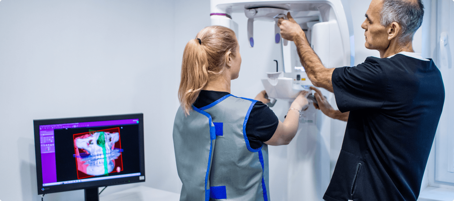
- Cone Beam Computed Tomography (CBCT)
All imaging procedures at Medicadent are conducted digitally, allowing for prompt consultations with specialists from other fields.
Types of dental radiology examinations offered at Medicadent clinic:
- Periapical X-rays
These are X-rays that cover only one tooth and its surrounding area. They are primarily used for specific tooth treatments, such as root canal therapy or dental implant placement. - Panoramic X-rays
This type of X-ray captures the upper and lower dental arches, allowing for a comprehensive view of the entire oral cavity. Panoramic X-rays are commonly used for dental diagnostics and orthodontic treatments.
- Cephalometric X-rays
Also known as lateral X-rays, as they present a profile view of the skull. They are primarily used in orthodontic treatments to assess the bite. - Cone Beam Computed Tomography (CBCT)
This examination is employed for detailed imaging of tissues and enables the detection of changes that may not be visible in regular X-rays. A distinctive feature of CBCT is the ability to obtain three-dimensional images, providing a view of structures from various angles. CBCT, also referred to as cone beam tomography, is utilized in periodontology, oral surgery, the diagnosis of temporomandibular joint disorders, and implantology.
Dental radiography plays a crucial role in various fields of dentistry. It is used in maxillofacial surgery to determine the position of impacted teeth within the bone. It is also performed prior to root canal therapy and during orthodontic treatment to assess bite irregularities and evaluate the effectiveness of procedures.